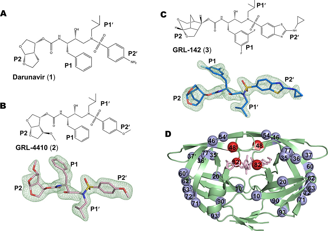
Figure 1. Compounds 1, 2, 3 and sites of mutation in PRS17 dimer.
A. Chemical structure of darunavir (1). B. Chemical structure and Fo-Fc omit map of 2 contour d at 3σ. C. Chemical structure and Fo-Fc omit map of 3 contour d at 3σ. D. PRS17 dimer in cartoon representation showing the sites of 17 mutations. The two active site mutations are shown as red spheres and the other mutations are blue spheres. Compound 3 bound at the active site is shown as pink sticks

