Fig. 2.

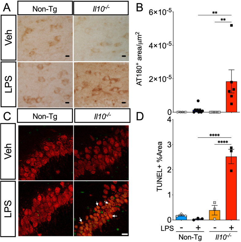
LPS-treated Il10−/− mice have increased neuronal p-tau and increased neuronal apoptotic cell death. Brain sections from Non-Tg or Il10−/− mice described above. A, B Immunohistochemistry (IHC) for endogenous pTau probed with AT180 antibody; 40× magnification; scale bar = 10 μm. A Representative sections from the dentate gyrus (DG) region of interest (ROI). B Quantification of AT180+ area. Data shown are mean ± SEM of area/μm2 from the standardized averages of three repeat trials; * p < 0.05, ** p < 0.01, *** p < 0.001; two-way ANOVA with Sidak’s multiple comparison test; n = 4–9 mice/group. C, D TUNEL immunofluorescence assay co-stained with NeuN antibody (arrows) to identify neurons in the Hp CA3 region; 63× magnification; scale bar = 10 μm. D Quantification of TUNEL+ area (green). Data shown are mean ± SEM of % area TUNEL+ cells in CA3 ROI; * p < 0.05, ** p < 0.01; *** p < 0.001; 2-way ANOVA; n = 3 mice/group; each individual data point shows an individual mouse sample
