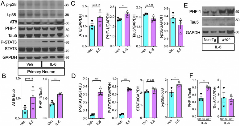
Fig. 6.
IL-6 stimulation increased p-tau in primary mouse neurons, in vitro. A WB of cell lysates from primary neuronal cell cultures directly treated with recombinant IL-6 protein (25 ng/mL) or Veh. B Quantification of pTau over total tau, AT8/Tau5, and PHF-1/Tau5 ratios. C Quantification of pTau (AT8/GAPDH and PHF-1/GAPDH) and total tau (Tau5/GAPDH) relative to loading control. D Quantification of (activated) phospho-STAT3 (P-STAT3) relative to (total) STAT3. Quantification of P-STAT3 and STAT3 relative to GAPDH. E, F WB of cell lysates of adult neurons from non-transgenic (Non-Tg) and IL-10-deficient mice (Il10−/−) show significantly higher PHF-1/Tau5 ratio in IL-10-deficient neurons treated with 25 ng/mL recombinant IL-6 compared to Non-Tg neurons treated with the same dose of IL-6. Data shown are mean ± SEM of IDV ratio; * p < 0.05, **p < 0.01; *** p < 0.001; unpaired t tests; n = 3 neuronal culture wells/treatment group

