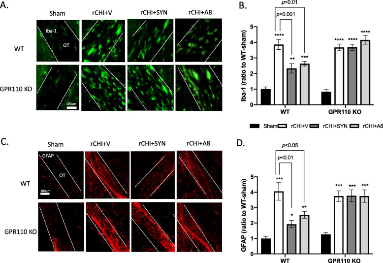
Fig. 4.
GPR110-dependent amelioration of rCHIMERA-induced glia cell activation by synaptamide and A8. A, C Representative immune-fluorescence micrographic images of Iba-1 (A) and GFAP (C) in the optic tract from WT and GPR110 KO mice at 1 week after injury (rCHI). WT and GPR110 KO mice were injected with A8 (1 mg/kg, i.p.) or synaptamide (5 mg/kg, i.p.) after each CHIMERA, and brains were collected for immunostaining at 7 days after the last injury. B, D Quantitative analysis showing significant suppression of Iba-1 (B) and GFAP expression (D) by the treatment with synaptamide or A8 compared to the vehicle-treated group (rCHI + V) in WT but not in GPR110 KO mice after rCHIMERA. The optic tract region (OT) is outlined with dashed lines. The data are expressed as mean ± SEM (n = 3). *p < 0.05, **p < 0.01, ***p < 0.001 vs. Sham-WT

