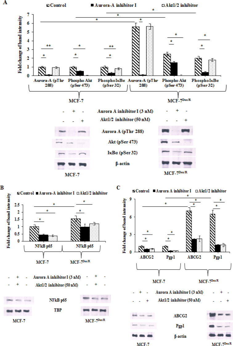
Figure 2.
Baseline Expressions and Corresponding Band Intensities of A. Aurora-A (pThr 288, Akt (pSer 473), IκBα (pSer 32), B. NFkB p65, C. ABCG2 and Pgp1 in parental and resistant sublines in presence or absence of either Aurora-A inhibitor I (3nM) or Akt1/2 inhibitor (50nM) for 6h followed by western blot analysis. To ensure equal protein loading β-actin/TBP was used. The experiments were repeated thrice. *p represents p<0.005 and **p represents p<0.01 in comparison to untreated cells. NFkB p65: Nuclear factor k B (p65 subunit); IκBα: nuclear factor of kappa light polypeptide gene enhancer in B-cells inhibitor alpha; ABCG2: ATP-binding cassette super-family G member 2; Pgp1: P-glycoprotein 1; TBP: TATA-Box binding protein

