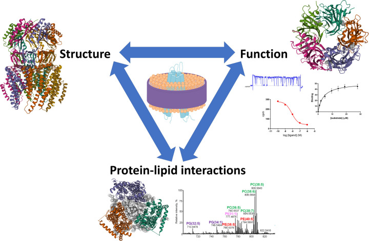
Figure 2. Polymer extraction of membrane proteins retains interactions between proteins and lipids which is important for both protein structure and function.
Side view structure of YnaI (PDB ID 6ZYD) [34].Top view structure of GlyR showing bound partial agonist taurine (space filling grey) (PDB ID 6PM0) [46], alongside representative images of various types of functional assays. Bottom view of AcrB trimer showing central lipid filled cavity (space filling grey) (PDB ID 6BAJ) [21], alongside a representative mass spectrum for lipids co-purified with a protein from yeast. Structural images made using Mol*[79].

