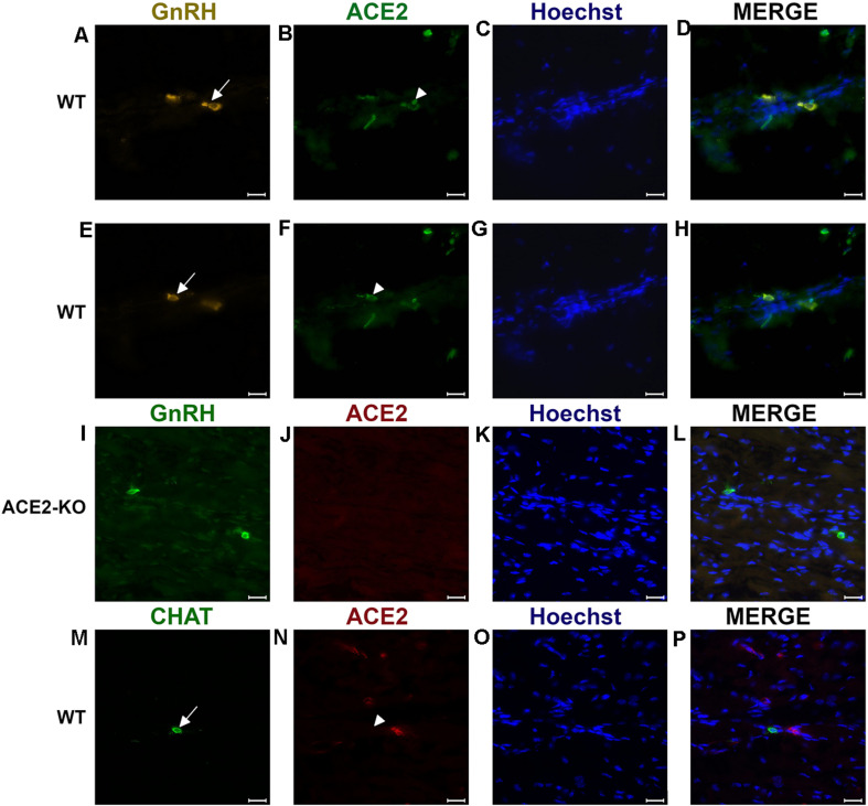
FIGURE 2.
Examples of double immunofluorescent labeling for nervus terminalis neuronal markers GnRH (A,E) or CHAT (M) and ACE2 (B,F,J,N) in the medial region adjacent to the olfactory bulbs as indicated in Figure 1. (A–H) Show slightly different focal planes to demonstrate the morphology of the two or three different neurons. Nuclei are stained with Hoechst 33258 (C,G,K,O). Merged images are shown in the last column (D,H,L,P). The neuronal somas labeled with GnRH (A,E) Are co-labeled with ACE2 (B,F) as shown after merging (D,H). GnRH positive cells in the ACE2 knock-out mouse (I) are not labeled with ACE2 (J). The majority of cholinergic neurons are not labeled with ACE2 (M,N), as quantified in Figure 3A. Control sections probed without primary antibodies or with control rabbit IgG had no detectable signal (not shown). Arrows and triangles indicate double-labeled neurons or lack thereof. Scale bars: 20 μm.

