FIGURE 4.

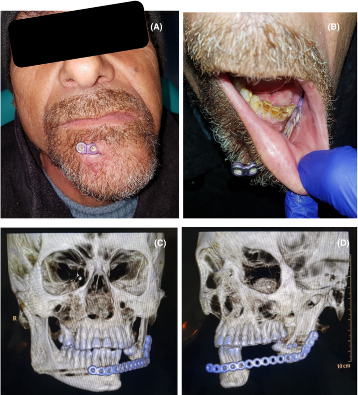
A‐B, Patient after 1 year of first surgery, intra‐ and extraoral view. C‐D, Computed tomography with broken 2.0 plate and lack of anterior fixation of the 2.4 plate

A‐B, Patient after 1 year of first surgery, intra‐ and extraoral view. C‐D, Computed tomography with broken 2.0 plate and lack of anterior fixation of the 2.4 plate