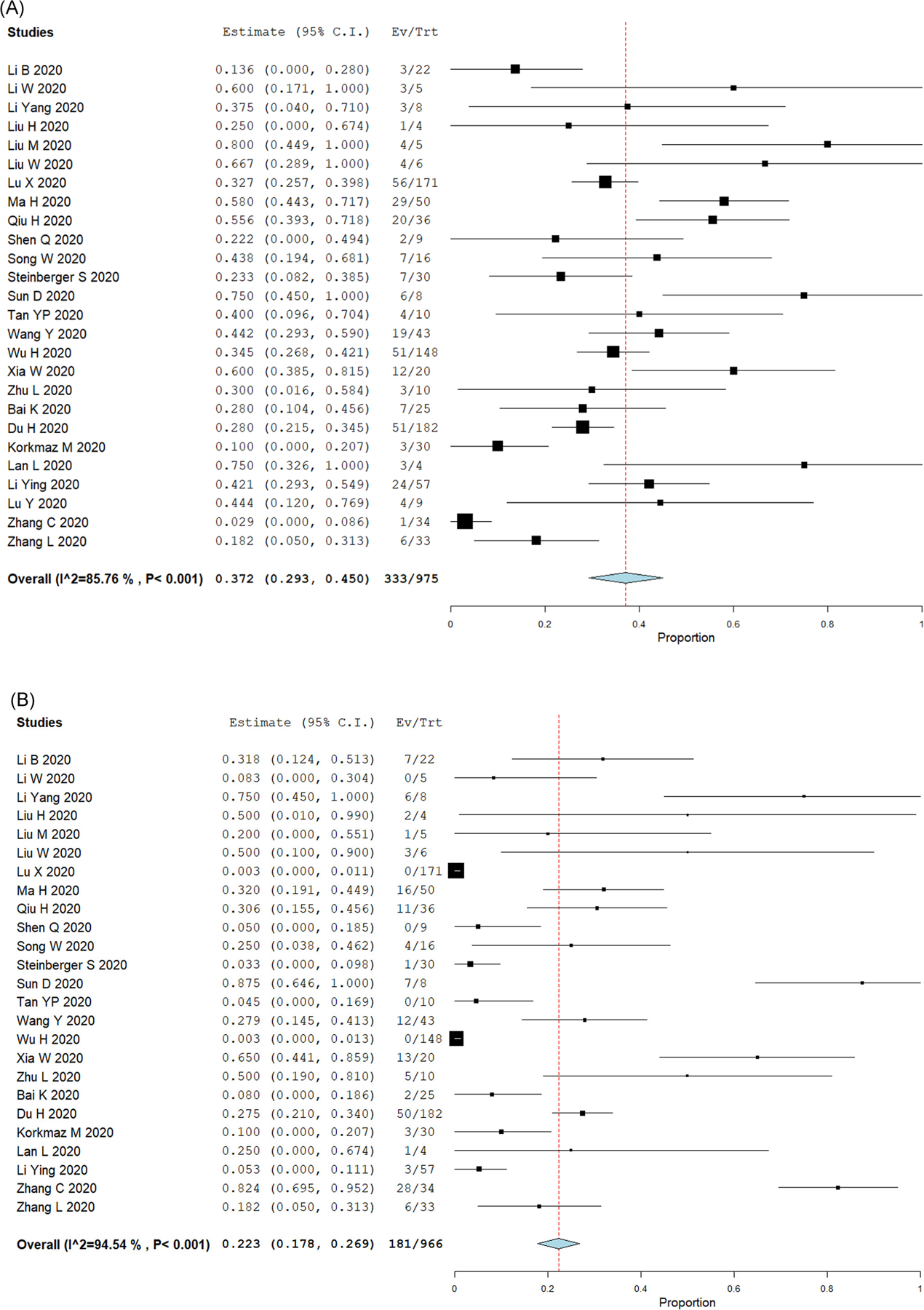
FIGURE 3.

Top lung imaging abnormalities in pediatric COVID-19 cases. Forest plots for single-arm meta-analysis of the pediatric COVID-19 studies reporting (A) ground-glass opacifications (GGO); or (B) consolidations or pneumonic infiltrates. Data presented as the 95% confidence interval (CI) of the proportion of subjects with these lung abnormalities in each study. COVID-19, coronavirus disease 2019 [Color figure can be viewed at wileyonlinelibrary.com]
