
FIGURE 4.
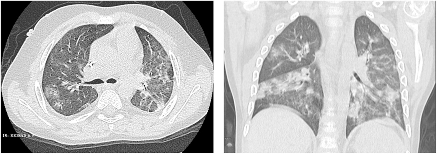
Typical radiological appearance of the most common abnormalities identified in pediatric COVID-19 cases. Chest CT of a 5-year-old male with PCR-confirmed SARS-CoV-2 infection. Images show multifocal scattered GGO and patchy consolidations with a peripheral distribution. There is no hyperinflation, increased perihilar markings, pleural thickening, pleural effusion, or bronchiectasis present. COVID-19, coronavirus disease 2019; CT, computerized tomography; GGO, ground-glass opacity; PCR, polymerase chain reaction; SARS-CoV-2, severe acute respiratory syndrome coronavirus 2