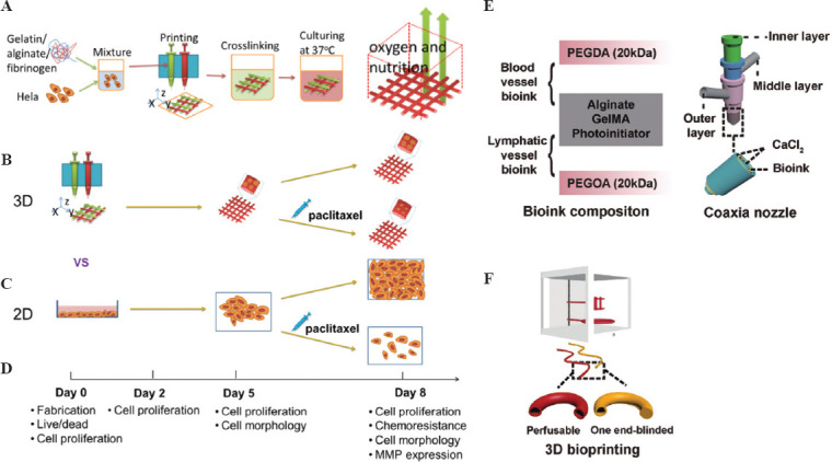
Figure 6.

(A) Schematic diagram of the method of 3D bioprinting tumor models with HeLa cells. (B) The plan of the 3D HeLa/hydrogel builds. (C) Both 3D HeLa/hydrogel constructs and 2D planar samples were incubated for 5 and 3 days with/without paclitaxel. The final results were compared. (D) Composition of bioink for bioprinting of blood vessels and lymphatic vessels. (E) Multi-layer coaxial nozzle design for bioink printing as well as cross-linking. (F) Two different hollow tubes were bioprinted using a perfusable hollow tube that mimics a blood vessel and an end-blind hollow tube that mimics a lymphatic vessel. (Adapted with permission from Yu Zhao et al, Biofabrication, 2014, 6 035001[67]) and (Adapted with permission from Cao X, Ashfaq R, Cheng F, et al., Adv Funct Mater, ©2019 WILEY-VCH Verlag GmbH & Co. KGaA, Weinheim[68]).
