Figure 1.
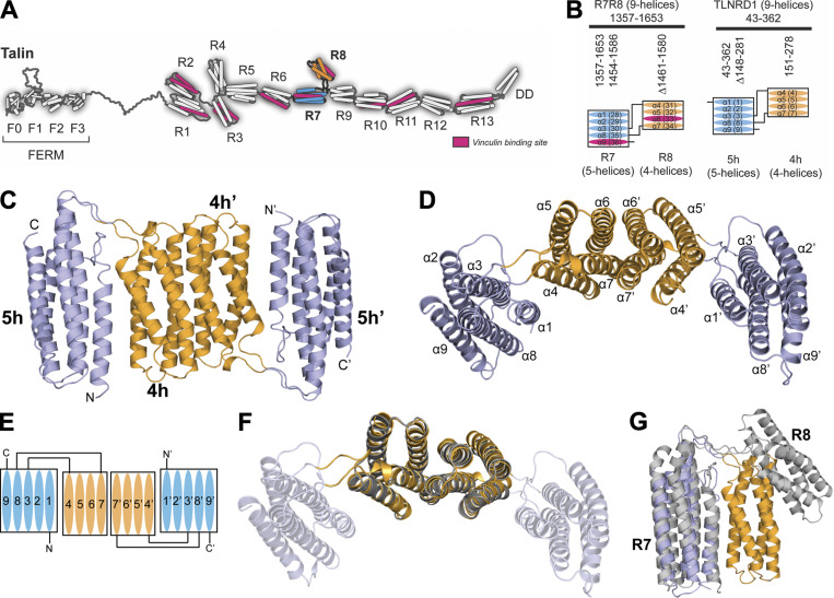
TLNRD1 is homologous to talin R7R8. (A) The domain structure of talin. Talin has an N-terminal head domain and a large rod domain comprised of 13 helical bundles, R1–R13 (Goult et al., 2013). The R7 (blue) and R8 (orange) domains are highlighted. The 11 VBSs are shown in pink. (B) Schematic of talin R7R8 (left) and TLNRD1 (right); the four-helix bundle (orange) is inserted into the loop between α3 and α8 helices of the five-helix bundle (blue). The VBS consensus sequence (Gingras et al., 2010) in talin R7 and R8 (pink) is absent from TLNRD1. (C) Crystal structure of TLNRD1-FL reveals an antiparallel, symmetric dimer. The domains of each monomer are labeled as 4h and 5h, and 4h’ and 5h’. (D) Top-down view of C with the helices labeled. The two four-helix bundles dimerize via the extensive interface between helices α6 and α7. (E) Schematic representation of the TLNRD1 dimer. (F) Overlay of the TLNRD1-4H structure (orange) on top of TLNRD1-FL (partially transparent) showing the dimerization interface is identical in both. (G) Overlay of one monomer of TLNRD1 (orange and blue) with talin R7R8 (gray; Protein Data Bank accession no. 2X0C). The individual domains are structurally homologous, but the orientation relative to each other is different, with talin R7R8 having a more open structure.

