Figure 3.
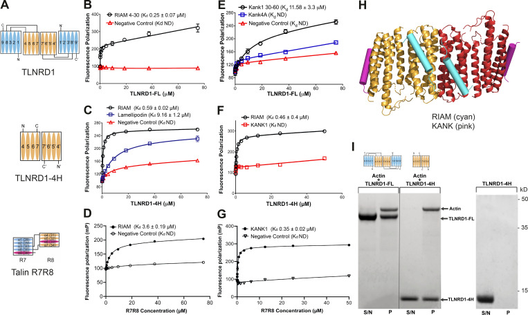
TLNRD1 and talin R7R8 both bind LD motif proteins and actin.(A) Schematics of TLNRD1-FL, TLNRD1-4H, and talin R7R8 domain structures. (B–G) TLNRD1 binds to LD motif–containing ligands. (B–D) Binding of fluorescein-labeled RIAM(4–30) peptide with (B) TLNRD1-FL, (C) TLNRD1-4H, and (D) talin R7R8 measured using a fluorescence polarization assay. The binding of lamellipodin (20–46; blue) is also shown. (E–G) BODIPY-labeled KANK1(30–60); wild-type (black) and 4A mutant (blue) peptides (E) binding to TLNRD1-FL, (F) not binding to TLNRD1-4H, and (G) binding to talin R7R8. ND, not defined. Experiments performed in triplicate. (H) Structural model of TLNRD1-FL bound to RIAM (cyan) and KANK (pink) LD motifs. (I) High-speed actin cosedimentation assay showing that both TLNRD1-FL and TLNRD1-4H interact with F-actin. TLNRD1-4H alone does not pellet. S/N, supernatant; P, pellet.

