Figure S3.
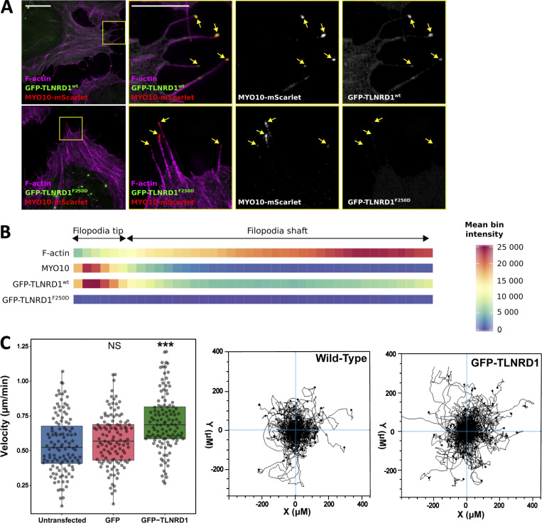
TLNRD1 localizes to the tip of MYO10 filopodia. U2OS cells expressing MYO10-mScarlet and GFP-TLNRD1 or GFP-TLNRD1-F250D were plated on fibronectin for 2 h, stained for F-actin, and imaged using SIM. (A) Representative maximum intensity projections are displayed. The yellow squares highlight regions of interest, which are magnified; scale bars: (main) 10 µm; (inset) 5 µm. (B) Heatmap highlighting the subcellular localization of F-actin, MYO10, TLNRD1, and TLNRD1-F250D within filopodia based on >360 intensity profiles (see Materials and methods for details). (C) Random 2D migration assay of U2OS cells plated on fibronectin and nontransfected or transiently expressing GFP or GFP-TLNRD1. GFP-TLNRD1 overexpression increases migration velocity in 2D (n = 120 cells from three independent repeats; ***, P < 0.001). Cell trajectories for nontransfected and GFP-TLNRD1–expressing cells are shown. P values were determined using a randomization test (see Materials and methods for details).

