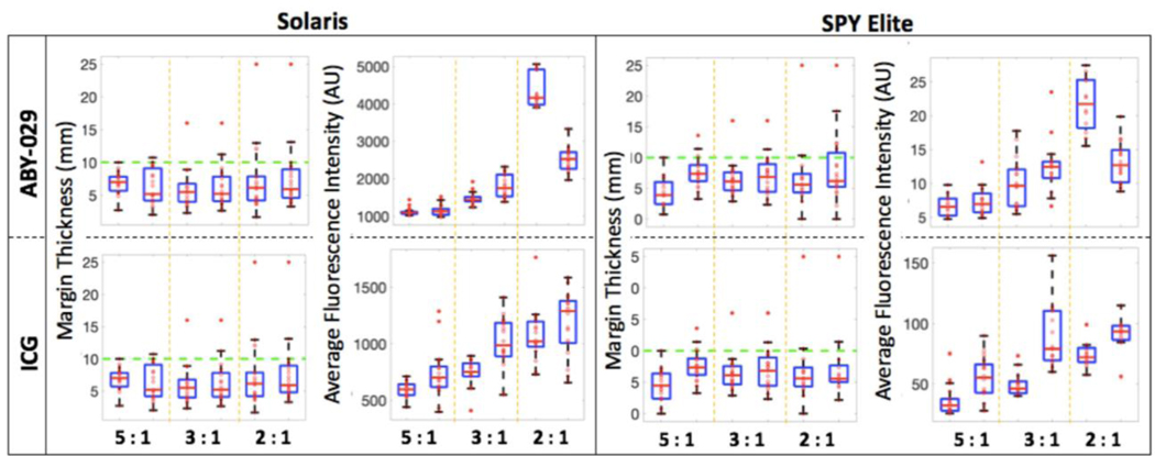
Figure 3.
Surgical phantom dissection data for ABY-029 and ICG. Margin thickness, and average fluorescence intensity from all six sides of the dissected inclusion measured both on the Solaris and the SPY Elite. The green dashed line on the margin thickness plots indicates the goal margin thickness of 10 mm.

