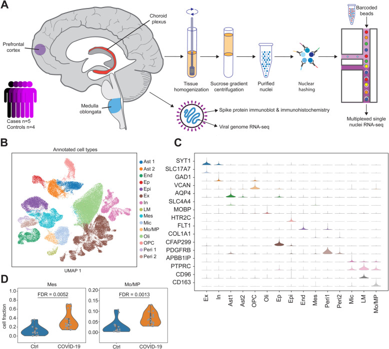
Fig. 1.
Droplet-based single-nucleus RNA sequencing in the dorsolateral prefrontal cortex (PFC), medulla oblongata (medulla), and choroid plexus (ChP) of 5 COVID-19 patients and 4 controls. A Experimental design. Frozen specimens of the human brain were dissected and subjected to a number of molecular assays, including single-nucleus RNA-sequencing (snRNA-seq), viral genome RNA-seq, and SARS-CoV-2 viral spike protein detection. B Uniform manifold approximation and projection visualization of annotated single-nucleus data (n = 68,557 barcodes). Colors show annotated cell types. C Distribution of canonical gene markers on annotated cell populations. The range of violins is adjusted by the maximum and minimum in each row. D Cell composition of mesenchymal cells and monocytes/macrophage in the choroid plexus stratified by case-control status. Only comparisons across tissues and cell types that survived false discovery rate correction are shown. Ast1 and Ast2 are the 2 groups of astrocytes. End, endothelial cells; Epi, epithelial cells; Ep, ependymal cells; Ex, excitatory neurons; In, inhibitory neurons; LM, lymphocytes; Mes, mesenchymal cells; Mic: microglia; Mo/MP, monocytes/macrophage; Oli, oligodendrocytes; Opc, oligodendrocyte progenitor cell. Peri1 and Peri2 are the two groups of pericytes

