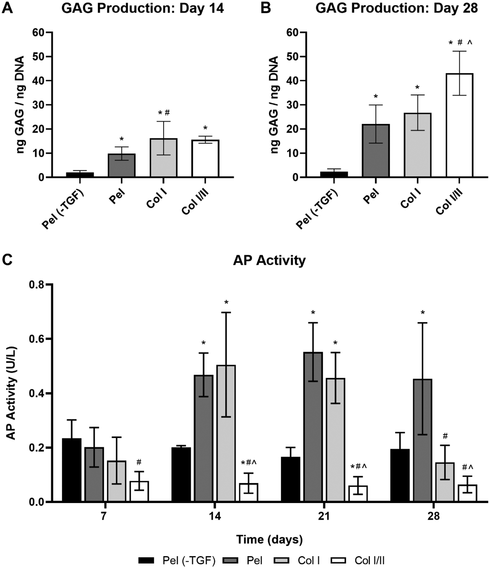
Figure 2. Collagen blend hydrogel increased GAG production when cultured with TGF-β and did not promote an increase in AP activity.

GAG/DNA ratio of the cell-hydrogel constructs and pellets after a (A) 14-day or (B) 28-day culture period. Values are expressed as mean ± SD (n = 5 or 6 for cell-gel scaffolds and pellets). A one-way ANOVA and Tukey’s post hoc test were performed within a timepoint (p < 0.05).* indicates a statistical difference in GAG production from pellets without TGF-β3 (Pel (-TGF)) within a timepoint, # indicates a statistical difference in GAG production from the pellets with TGF-β3 (Pel) within a timepoint, and ^ indicates a statistical difference in GAG production from the collagen type I hydrogels (Col I) within a timepoint. (C) AP activity of rabbit MSCs over time in removed culture medium at 7, 14, 21, or 28 days (n = 8 or 9 for Col I hydrogels, Col I/II hydrogels, and pellets with TGF-β3 (Pel) and n = 3 for pellets without TGF-β3 (Pel (-TGF))). A mixed-effect model and Tukey’s post hoc test were performed (p < 0.05). * indicates a statistical difference in AP activity from pellets without TGF-β3 (Pel (-TGF)) within a timepoint, # indicates a statistical difference in AP activity from the pellets with TGF-β3 (Pel) within a timepoint, and ^ indicates a statistical difference in AP activity from the collagen type I hydrogels (Col I) within a timepoint.
