
Figure 5. Col I/II hydrogel promoted cartilage matrix production in vivo.
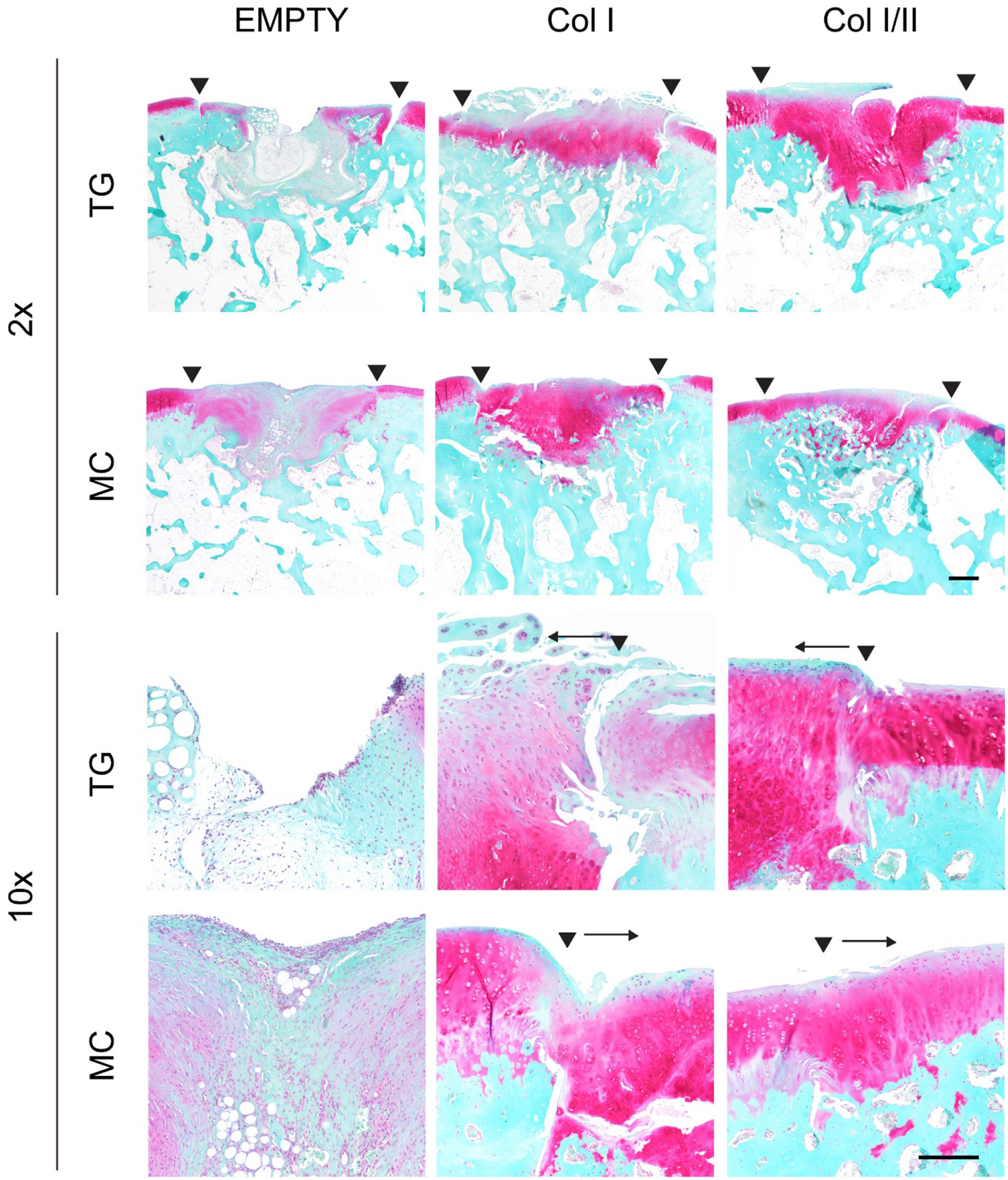
SOFG staining of repaired cartilage left empty or filled with either a Col I or Col I/II hydrogel in the trochlear groove (TG) or the medial condyle (MC) at two different magnifications (2x with a scale bar of 500 μm and 10x with a 200 μm scale bar). The Col I/II hydrogels had dark pink staining, which indicated the presence of proteoglycans. The two arrowheads indicate the edges of the repair tissue. A single arrowhead with an arrow indicates the edge of the repair tissue and the direction of the repair tissue, respectively.