Fig. 1.
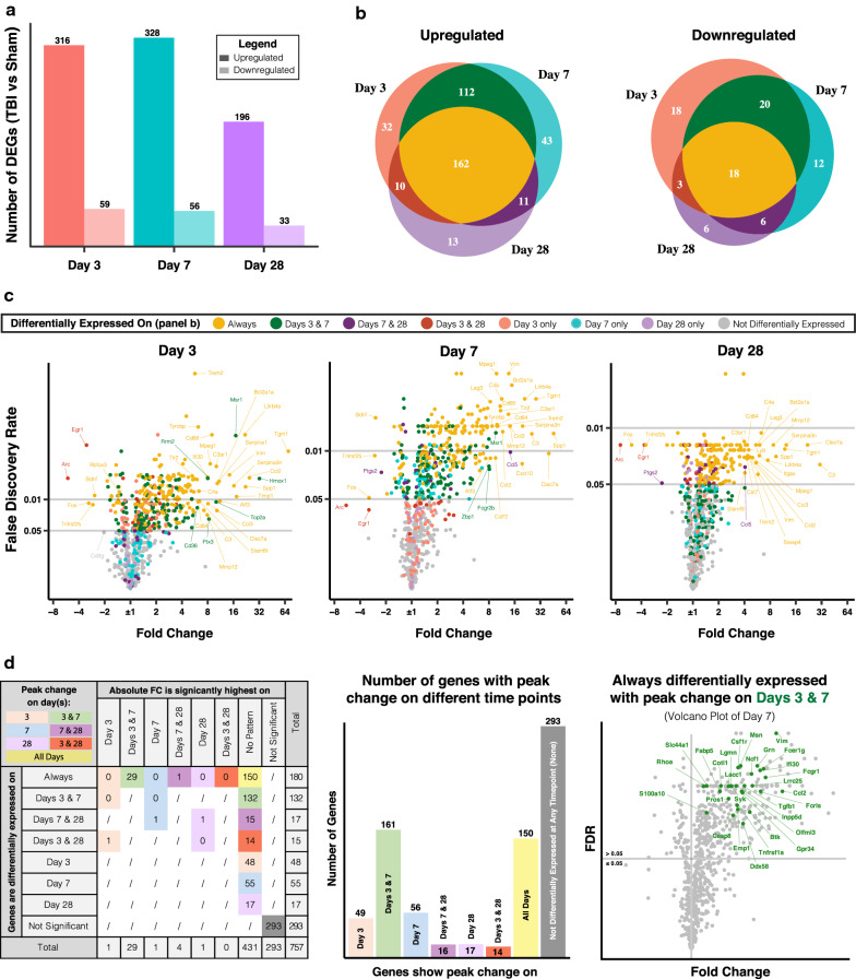
Patterns of neuroinflammatory gene expression in TBI. a Number of upregulated and downregulated genes in injured brains at days 3, 7, and 28 after TBI compared to 12-week-old uninjured brains (using NanoString Neuroinflammation panel). b Common changes in gene expression between time points color coded by the time point(s) of differential expression. c Volcano plots of differentially expressed genes per time point, color coded as in panel b, and showing labeled examples of the most dysregulated genes. d Analysis of time points of peak change in expression per gene (color legend) based on statistical difference in fold change (columns) between time points of differential expression (rows). Genes that showed no significant differences were considered to show peak change at all time points of differential expression. The volcano plot on the right shows an example of 29 consistently upregulated genes that had peak expression on days 7 and 28 post injury compared to day 3 post injury. False discovery rate (FDR) was computed for all genes per comparison to determine statistical significance. N = 3 per group

