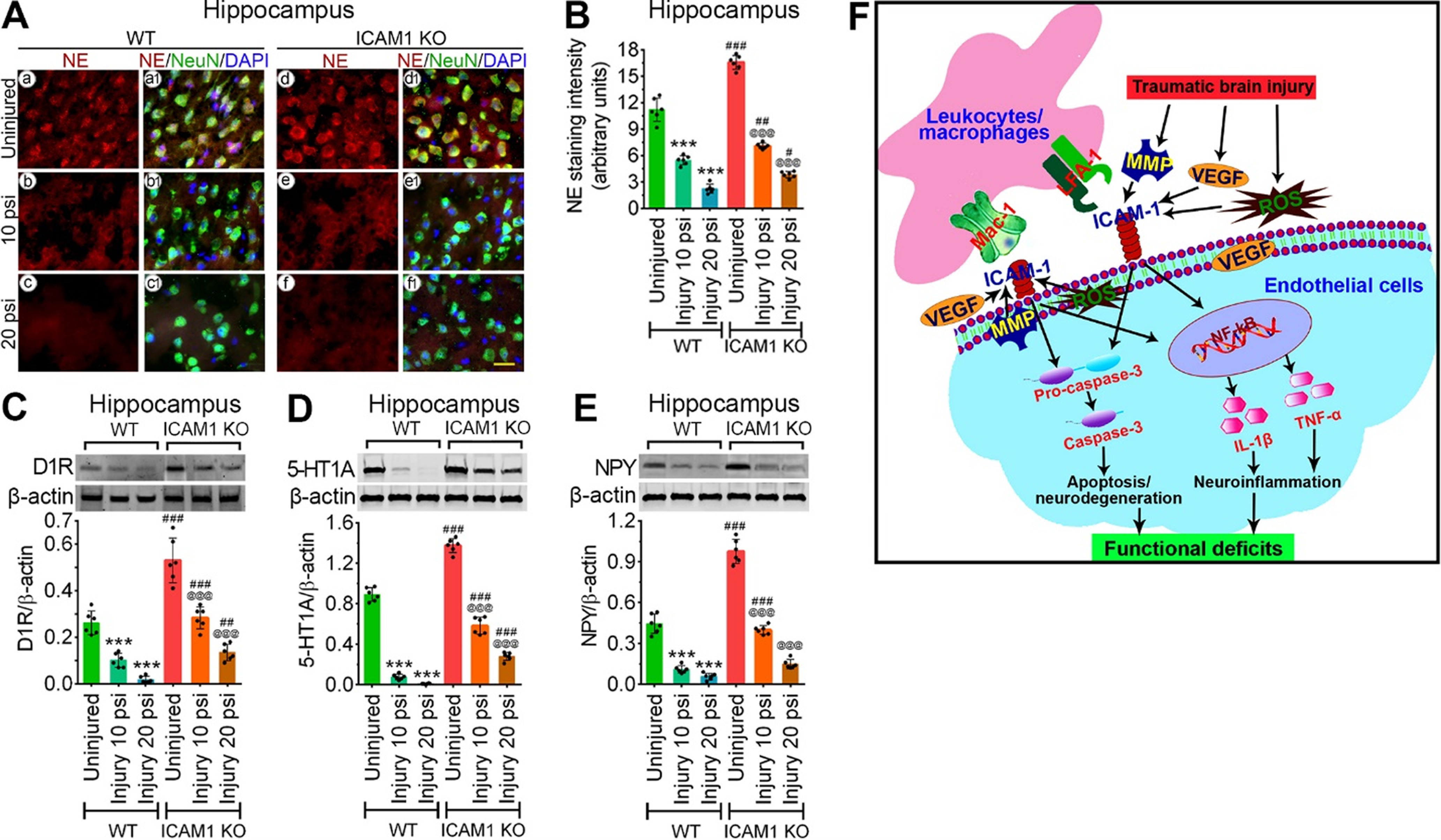
Figure 8.

ICAM-1 reduces neurotransmitters expression that reflects in sensorimotor deficits and psychological stress after TBI. A, Immunofluorescent staining of NE (red) in the hippocampus area of WT and ICAM-1−/− mice after 10 and 20 psi FPI and merged with NeuN (green) and DAPI (blue). Scale bar: 20 μm (all panels). B, Quantification of NE staining in the hippocampus area of uninjured, 10 and 20 psi FPI WT and ICAM-1−/− mice using ImageJ software (n = 6/group). C–E, Western blot analysis of 5-HT1AR (C), DAD1R (D), NPY (E) and β-actin in the tissue lysates of hippocampus of WT and ICAM-1−/− mice 48 h after 10 and 20 psi FPI. The bar graph with dot plots shows the quantification of 5-HT1AR (C), DAD1R (D), NPY (E) versus β-actin (n = 6/group). F, Schematic presentation of the findings. All values are expressed as mean ± SD two-way ANOVA followed by Bonferroni post hoc tests. Statistically significant ***p < 0.001 versus WT uninjured group; @@@p < 0.001 versus uninjured ICAM-1−/− group; #p < 0.05, ##p < 0.01, ###p < 0.001 versus WT corresponding injury groups; ns = non-significant. NE, norepinephrine; 5-HT1AR, 5-HT 1A receptor; DAD1R, DA D1 receptor; NPY, neuropeptide Y.
