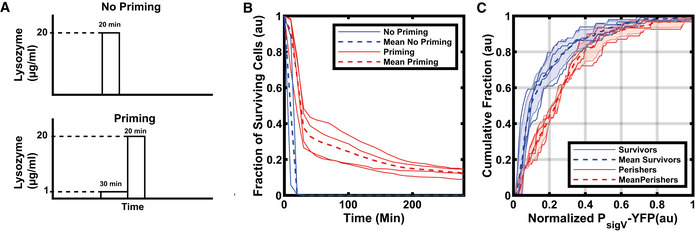
Figure 2. Rapid activation of σV after a first stress application increases survival after a second higher stress.

-
ASchematic of lysozyme application. Cells are either exposed directly to a high concentration of lysozyme (20 µg/ml) for 20 min (top) or exposed first to a short (30 min) lysozyme priming stress (1 µg/ml) before exposure to the higher concentration (bottom).
-
BA priming (30 min) stress of 1 µg/ml lysozyme followed by the high lysozyme stress (20 µg/ml) improves survival. The solid blue lines are the biological repeats for the no priming experiment (n = 2) whereas the solid red lines are the biological repeats for the priming experiment (n = 4). (Total number of cells shown for No Priming, N = 2,013 and Priming, N = 4,937).
-
CSurviving cells have higher P sigV ‐YFP levels after the initial priming stress (1 µg/ml) than perishing cells. The cumulative distributions were normalized by their maximum σV activity and baseline subtracted. Each dashed line is the mean of experiments from n = 4 biological repeats. The shaded areas correspond to the mean ± s.d. For more information on the number of repeats and cell numbers, please see the supplementary text.
Data information: For more information on the number of repeats, please see Appendix Table S3.
