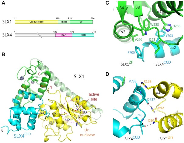
Figure 1.
Structure of the yeast SLX1–SLX4 complex. (A) Schematic drawing of domain structures of yeast SLX1 and SLX4 proteins. (B) 1.45 Å structure of the SLX1–SLX4CCD complex shown in a cartoon representation, with the Uri/GIY-YIG) domain, zinc finger (ZF) domain and the linker helix of SLX1 colored, yellow, green and pale green, respectively, and the SLX4 CCD domain is colored cyan. α-helices and β-strands in each domain are numbered consecutively and labeled. The grey spheres indicate zinc ions in the ZF domain. The conserved residues in the catalytic active site, which is indicated by dashed-line circle, are shown in a stick representation. (C) Mostly hydrophobic interaction between the ZF domain of SLX1 and the CCD domain of SLX4, and involved residues are displayed as sticks. (D) Chiefly polar interaction mediates packing of the Uri domain of SLX1 and the CCD domain of SLX4. Dashed lines indicate intermolecular hydrogen bonds between the involved residues.

