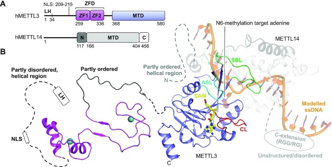
Figure 2.
Structure of the human METTL3:METTL14 complex. (A) Schematic outline of the human METTL3:METTL14 (hMETTL3:hMETTL14) complex protein domains. Crystallized parts are indicated in boxes. (B) Structure of human METTL3 ZFD (light pink) (PDB 5YZ9), SAM-bound (yellow) MTD (blue) (PDB 5IL1) complexed with METTL14 covering the N-extension (black) and MTD (grey) of human METTL14 (PDB 5IL1). The sections not included in the structures are shows as dotted lines, and their structure/order state is indicated. The catalytic loop (CL, red), the active site loop (ASL, cyan) and the substrate binding loop (SBL, green) are shown for METTL3. Single-stranded DNA (ssDNA) (orange) has been modelled from the DNA-bound m6A DNA MTase EcoP15I (PDB 4ZCF).

