Figure 5.
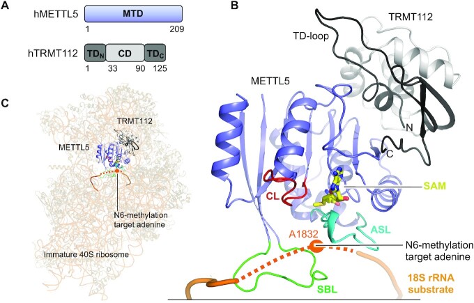
Structure of the human METTL5:TRMT112 complex. (A) Schematic outline of the human METTL5:TRMT112 (hMETTL5:hTRMT112) complex protein domains. Crystallized parts are indicated in boxes. (B) Structure of the SAM-bound (yellow) full-length, human METTL5 (blue) (PDB 6H2V) bound to human TRMT112 (grey/black) (PDB 6H2U). The active site loop (ASL, cyan), catalytic loop (CL, red) and substrate binding loop (SBL, green) are shown. Substrate 18S rRNA (dark orange) is modelled from positioning of METTL5:TRMT112 on the immature 40S ribosome (PDB 6G53). The immature ribosome structure lacks U1830-A1835 from the rRNA (not build), which is here instead indicated as a dotted line and the target base (A1832) is indicated as an orange dot. (C) Positioning of METTL5:TRMT112 into unoccupied density on the surface of the immature 40S ribosome (PDB 6G53). The unbuild part of the rRNA (U1830-A1835) is shown as a dotted line and the target base (A1832) is indicated as an orange dot.

