Figure 7.
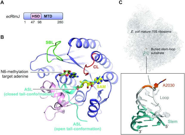
Structure of E. coli RlmJ. (A) Schematic outline of the E. coli RlmJ (ecRlmJ) protein domains. Crystallized parts are indicated in boxes. (B) Structure of the ecRlmJ SAM-bound (yellow) MTD (blue) with the inserted HSD (pink) (PDB 4BLV), crystallized with a closed tail-conformation of the active site loop (ASL, cyan). The ASL is further shown from apo RlmJ (PDB 4BLU) in the open tail-conformation. The target adenine (dark green) is modelled from RlmJ co-crystallised with a bisubstrate molecule (BA4) (PDB 6QDX, chain B). The catalytic loop (CL, red) and substrate binding loop (SBL, green) are shown. (C) The mature E. coli 70S ribosome with an insert highlighting the RlmJ substrate stem-loop structure from 23S rRNA with target adenine (A2030, dark orange) placed in the loop (grey) followed by a stem (teal) (PDB 7K00).

