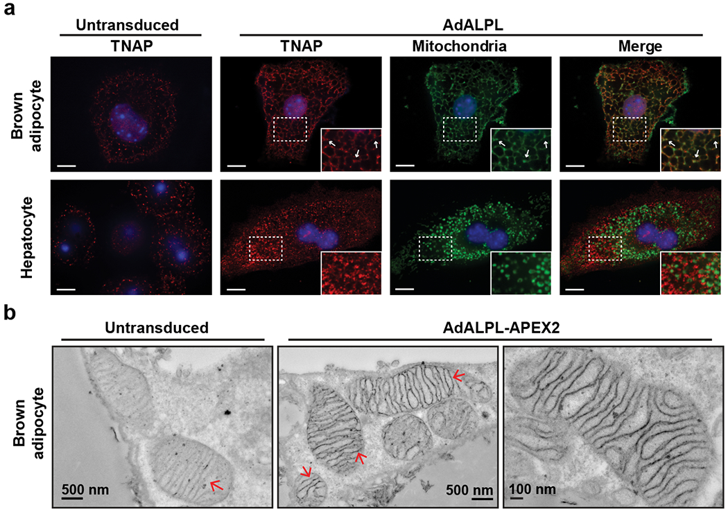
Figure 2. TNAP targets mitochondria in brown adipocytes.

(a) Confocal fluorescence images showing subcellular localization of ectopically expressed TNAP in immortalized brown adipocytes and primary hepatocytes. The insets show an enlarged region of the image outlined by the dotted box. Arrows denote selected signals of TNAP that colocalize with mitochondria signal. Anti-TNAP (Red) and anti-HSP60 (Green) were used to visualize TNAP and mitochondria, respectively. Scale bar: 10 μm.
(b) Transmission electron microscope images showing the detailed localization of TNAP-APEX2 in primary mature brown adipocytes. Arrows denote the structures of mitochondrial inner membrane.
