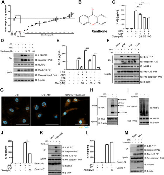
FIGURE 1.

Xanthone dose‐dependently inhibits NLRP3 inflammasome activation with no effect on priming step. (A) Unbiased screening of natural NLRP3 inhibitor using LPS‐primed peritoneal macrophages. IL‐1β level of LPS+ATP group (positive control, P.C) was set as 1.0. (B) Structure of xanthone. (C and D) LPS‐primed peritoneal macrophages treated with different doses of xanthone 2 h before ATP challenge. Supernatants (SN) and cell extracts (lysate) were analyzed by immunoblotting (D), IL‐1β (C) secretion was determined by ELISA. (E and F) LPS‐primed peritoneal macrophages treated with 50 μM xanthone for 2 h, followed by stimulation with ATP, Nigericin (N), aluminum salts (Alum). Supernatants (SN) and cell extracts (Lysate) were analyzed by immunoblotting (F). Supernatants were also analyzed by ELISA for IL‐1β (E). (G and H) Representative immunofluorescence images of ASC speck formation of LPS‐primed BMDMs with indicated treatment (G), and ASC oligomerization in cross‐linked cytosolic pellets analyzed by immunoblotting in (H). Scale bars, 20 μm. (I) Semi‐denaturing detergent agarose gel electrophoresis (SDD‐AGE) detection of NLRP3 oligomerization. (J–M) LPS‐primed peritoneal macrophages treated with 50 μM xanthone before 100 μM Imiquimod challenge (J and K) or substitution of K+‐free medium challenge for 30 min (L and M). Supernatants (SN) and cell extracts (Lysate) were analyzed by immunoblotting (K and M), IL‐1β (J and L) secretion was determined by ELISA. *p < 0.05, **p < 0.01, ***p < 0.001, ****p < 0.0001, two‐tailed unpaired Student's t‐test. Data are the mean ± SD
