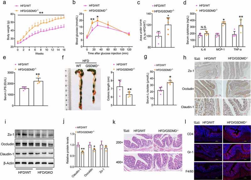
Figure 3.

GSDMD Restrains HFD Feeding-induced Systemic Endotoxemia
WT and GSDMD−/- mice were fed a HFD for 16 weeks.(a) Body weight. (b) Glucose tolerance test. (c) Area under curve of Figure 3b. (d) Serum levels of proinflammatory cytokines, including IL-6, MCP-1 and TNF-α. (e) Serum levels of LPS. (f) Morphological analysis of the colons. (g) Serum D-lactate levels. (h) IHC analysis of the colonic expression of permeability-associated proteins, including Zo-1, Occludin and Claudin-1 (Scale bars, 50 μm). (i) Protein levels of Zo-1, Occludin, and Claudin-1 in the colons. (j) Densitometric analyses of Figure 3i. (k) H&E staining of the colonic sections (Scale bars, 50 μm). (l) IF analysis of inflammatory markers, including CD4, Gr-1, and F4/80 (Scale bars, 50 μm). For all the panels, n = 5 mice/group. All the values are presented as the mean ± SD. The data were analyzed by using one-way ANOVA followed by Fisher’s LSD post hoc test. *P < .05, **P < .01 compared to the HFD-fed WT group.
