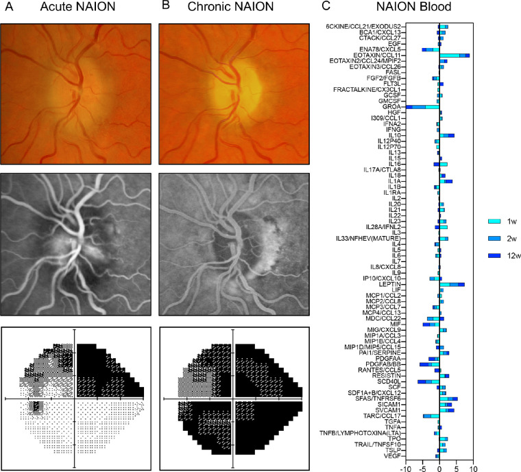
Figure 1.
NAION description and blood profiling over time. A presentative right eye with non-arteritic ischemic optic neuropathy (NAION) in a 70-year-old male patient baseline, acute NAION (A), and chronic NAION (B). From the top row to the bottom row, the figure shows the fundus photo, optical coherence tomography and angiography, and grayscale displays of Humphrey visual field. (C) Blood multiplex immunoprofiling at 1 (turquoise), 2 (sky blue), and 12 (dark blue) weeks after NAION onset. Ratio to 9 controls was log2-scaled.

