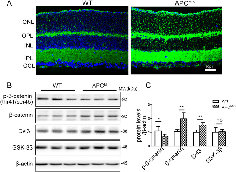
Figure 2.
Activation of the canonical Wnt signaling pathway in APCMin mice. (A) Ocular sections from wt and APCMin mice on P84 were stained with the anti-β-catenin antibody (green), and the nuclei were counterstained by DAPI (blue). (B) The same amount of eyecup proteins (100 µg) was blotted separately with antibodies for phosphorylated-β-catenin, total β-catenin, Dvl3, and GSK3β. (C) Semi quantified by densitometry and normalized by β-actin levels. The Figure is a summary of two independent experiments with n = 6 eyes per group Student t-test. Data are presented as mean ± SEM, *P < 0.05, **P < 0.01. ns, no difference.

