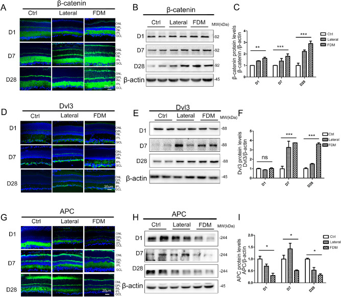
Figure 3.
The canonical Wnt pathway is progressively activated during myopia development. (A, B) β-catenin expression was evaluated by immunostaining and western blot analysis in FDM and normal control groups following day 1, day 7, and day 28 of treatment. (C) β-catenin protein levels were semi quantified with densitometry and normalized by β-actin levels. (D, E) Dvl3 expression was evaluated by immunostaining and Western blot analysis in FDM and normal control groups following day 1, day 7, and day 28 of treatment. (F) Dvl3 protein levels were semi quantified with densitometry and normalized by β-actin levels. (G, H) APC expression was evaluated by immunostaining and Western blot analysis in FDM and normal control groups following day 1, day 7, and day 28 of treatment. (I) APC protein levels were semi quantified with densitometry and normalized by β-actin levels. Data are presented as mean ± SEM, n = 10, *P < 0.05, **P < 0.01, compared with the normal group and lateral eye of FDM.

