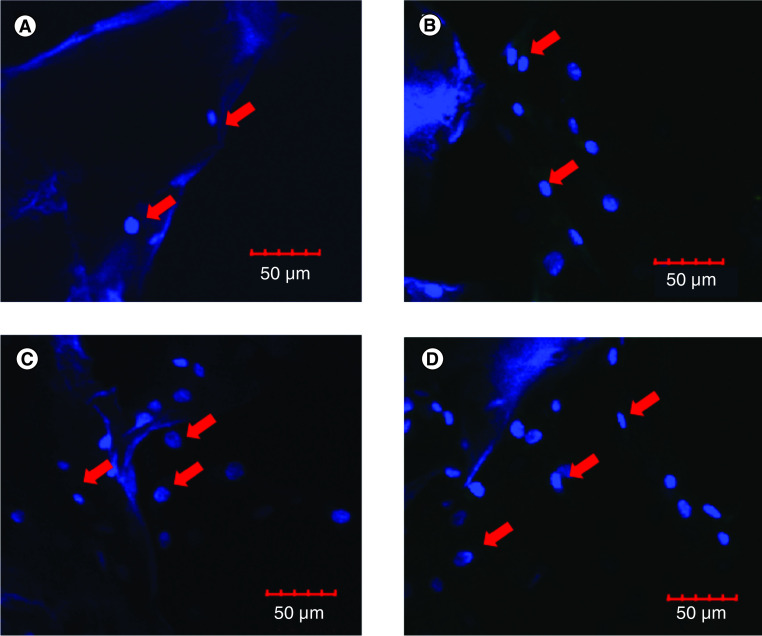
Figure 4. . Visualization of cell penetration in silk fibroin scaffold by using confocal laser scanning microscopy.
(A) hADSCs cultured on scaffold for 7 days, upper surface of scaffold. (B) hADSCs cultured on scaffold for 21 days, upper surface of scaffold. (C) hADSCs cultured on scaffold for 7 days, lower surface of scaffold. (D) hADSCs cultured on scaffold for 21 days, lower surface of scaffold. The red arrows indicate nucleus.
hADSC: Human adipose-derived stem cell.

