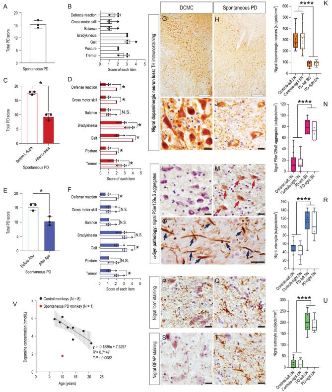
Figure 1.
Classic Parkinsonian symptoms and pathologic hallmarks of the spontaneous PD monkey (#06103). (A) Quantified by the improved Kurlan scale, the monkey had a total PD score above 15 out of a maximum of 20, demonstrating severe PD symptoms. (B) The seven items’ scores that constitute the improved Kurlan scale were all above zero, revealing the monkey had all the diagnostic PD symptoms. The total PD score was the sum of the seven individual item scores. (C) The total PD score of the monkey was reduced significantly from 17.7 to 9.3 after a classic PD drug L-dopa treatment, indicating a significant positive response to the treatment. (D) The scores of the seven individual items were all reduced after the L-dopa treatment, and the reduction of six items out of the seven were significant. Both C and D provide strong evidence for pharmacological validation. (E) The total PD score of the monkey was reduced significantly from 15.3 to 10.3 after the apomorphine (Apo, an agonist of dopamine D1 receptor) treatment, indicating a significant improvement of PD symptoms by dopamine D1 receptor activation. (F) The scores of the seven individual items were all reduced after Apo treatment except for gross motor skill, and three out of the seven items were significant. These data further confirmed the above results from the L-dopa treatment. The data were quantified from three video clips of each experimental condition and presented as mean ± SD. Non-parametric statistics (Mann-Whitney test) were used (*, P < 0.05). (G and H) Under lower resolution images (4×), nigral TH immunostaining of the death-condition-matched control (DCMC, #071809) and spontaneous PD monkey indicated the overall nigral dopaminergic neuron loss of the spontaneous PD monkey: fewer nigral dopaminergic neurons that were lightly stained survived in the spontaneous PD monkey compared with the DCMC. (I and J) Under higher resolution images (40×), nigral TH immunostaining of the spontaneous PD monkey showed obviously less dopaminergic neurons with weakly stained somata and fewer neurites compared with that of the DCMC. (K) Quantitative analysis demonstrated that the nigral dopaminergic neuron number of the spontaneous PD monkey was about 70% less compared with that of the controls (****, P < 0.001). (L and M) PSer129αS immunostaining of the SNs revealed that there were more PSer129αS aggregates (red arrows) in the spontaneous PD monkey compared with the DCMC. (N) The number of PSer129αS aggregates in the spontaneous PD monkey's SN was five times higher (****, P < 0.001). (O) PSer129αS aggregates in the nigral neuron's processes were found in the spontaneous PD monkey (blue arrows). (P and Q) Iba1 immunostaining of the monkeys’ SNs revealed that there were more activated microglia in the spontaneous PD monkey compared with the DCMC. (R) The number of activated microglia in the spontaneous PD monkey's SN was 2.5 times larger than the controls (****, P < 0.001). (S and T) GFAP immunostaining in the monkeys’ SNs revealed that there were more activated astrocytes in the spontaneous PD monkey compared with the DCMC. (U) The number of the activated astrocyte in the spontaneous PD monkey's SN was 12.5 times larger than the controls (****, P < 0.001). Scale bars: hollow black: 200 μm; solid black: 20 μm. Data in K, N, R and U are median with minimum to maximum. Non-parametric statistics (Mann-Whitney test) was used. (V) Lower cerebrospinal fluid (CSF) dopamine level of the spontaneous PD monkey compared to the eight normal controls. The dopamine level was measured by a high-pressure liquid chromatography. Black dots represent dopamine concentrations of the eight normal control monkeys. The black line is the regression curve, and the gray shadow represents the 95% confidence interval of the control monkeys’ CSF dopamine concentration. The red dot represents the CSF dopamine concentration of the spontaneous PD monkey, which is located way outside the 95% confidence interval. *, P < 0.05; **, P < 0.01 and ****, P < 0.001.

