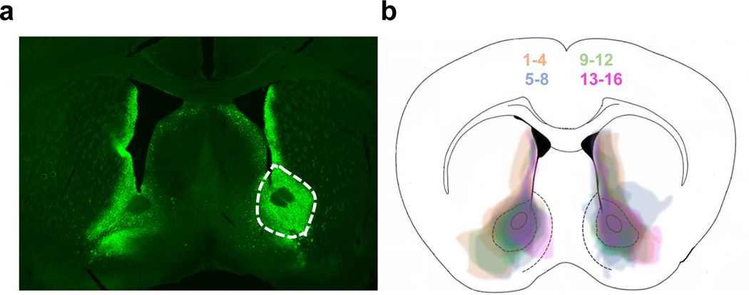
Figure 1. Confirmation of viral spread.
(a) Coronal section showing D2R-mVenus expression in the NAc of a Drd2-Cre mouse. (b) Superimposed traces of viral spread from coronal sections at ~1.0 mm anterior to bregma for all 16 D2ROENAcInd mice injected with the AVV1-hSyn-DIO-D2R-mVenus into the NAc. Mice are shown in 4 different colors (4 mice/color) for better visualization of viral spread.

