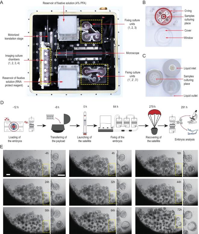
Figure 1.
In vitro development of mouse pre-implantation embryos in space. (A) The embryonic culture incubator used in space experiments. The incubator consists of four imaging culture chambers (indicated by the yellow dotted line and the numbers 1, 2, 3 and 4), two groups of culture fixation units (yellow dotted lines), a microscope (red arrow), and two module reservoirs of fixative solutions (red arrow). This experimental apparatus provides temperature stability inside the incubator, the ability to replace culture medium and automatic image acquisition. (B) The imaging culture chamber was filled with gas-saturated medium, and it consisted of a sample culture cavity with a red O-ring and a cover with a window. (C) The fixation culture unit is a cylindrical perfusion chamber with a top part of the chamber, main body and bottom part of the chamber. (D) Timeline of the SJ-10 satellite space mission showing the time points for embryos loading, payload transferring, embryos fixation, sample recovery and arrival to the laboratory. (E) Representative time-lapse images of embryonic development in imaging culture chambers during spaceflight, with highlighted images showing key stages of pre-implantation development. Scale bars, 100 μm.

