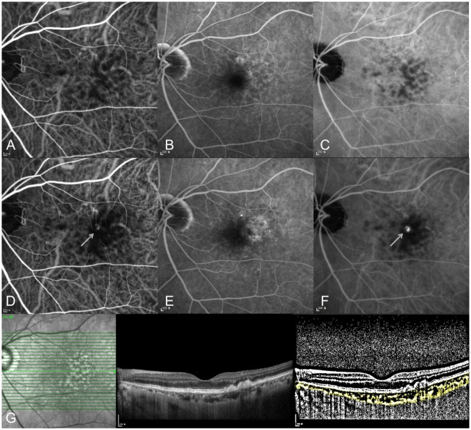
Fig 1. A 72-year-old woman with right eye type 3 neovascularization.
(A-C) At initial diagnosis, no neovascular lesion was detected in her left eye at early-to-late phase on indocyanine green and fluorescein angiography. (A) early phase indocyanine green angiography, (B) late phase and fluorescein angiography, (C) late phase indocyanine green angiography. (D-F) Ten months later, type 3 neovascularization progressed in her left eye. The arrow indicates the neovascular lesion on indocyanine green angiography. (D) early phase indocyanine green angiography, (E) late phase and fluorescein angiography, (F) late phase indocyanine green angiography. (G) OCT B-scan and binarized image to measure the choroidal vascularity index (CVI) in the left eye at initial diagnosis of type 3 neovascularization in the right eye.

