Figure 5.
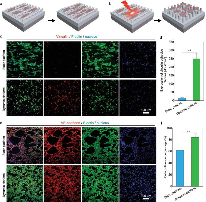
Focal adhesion and intercellular junction of human ECs on different platforms with either static topography or dynamic topography. Schematic illustrations of cell functions directed by either static platform (a) or dynamic platform (b). (c) Representative fluorescence images of HUVECs on different platforms on day 3 of culturing, showing similar cell density (indicated by DAPI-stained nucleus) yet distinctly different results in cytoskeleton organization and cell focal adhesion. (d) Statistical analyses on the expression of vinculin adhesive plaques for the HUVECs grown on different platforms on day 3 of culturing. (e) Representative fluorescence images of HUVECs on different platforms on day 7 of culturing, exhibiting both abundant expression of VE-cadherin and similar cell density (indicated by DAPI-stained nucleus), yet different conditions in cytoskeleton organization and cell-confluence rates. (f) Statistical analyses on the cell-confluence percentages of HUVECs grown on different platforms on day 7 of culturing, demonstrating a statistically significantly higher confluence rate of HUVECs on the platform with dynamic topography than that on the platform with static microgroove-array topography.

