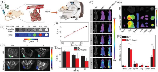
Figure 5.
(A) Schematic illustration of EMM nanocomposites effectively accumulated at a tumor site under magnetic targeting for T2-weighted MR imaging. (B) The T2-weighted image, map and (C) the transverse relaxivity r2 of EMMs. (D) The in vivo T2-weighted images and (E) normalized T2 signal change of 4T1 tumor-bearing mice after intravenous injection of MM nanocomposites in the absence and presence of magnetic targeting. (F) The fluorescence images of 4T1 tumor-bearing mice after intravenous injection of DiR-loaded MMs (MMDiR) at 1, 2, 4, 10 and 24 h. (G) Fluorescence images and (H) quantitative results of DiR fluorescence intensity in major organs and tumor.

