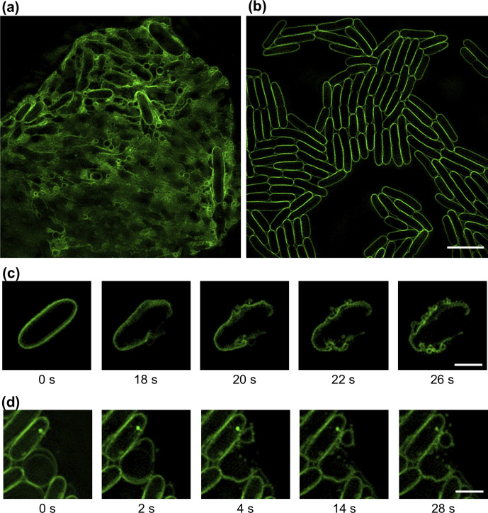
Fig. 3.
Explosive cell lysis generates MVs in E. coli . (a, b) 3D-SIM images of E. coli MG1655 (a) infected with phage T7 or (b) treated with phage buffer control. Images of E. coli infected with T4 phage were similar to (a). (c, d) Time-lapse image sequences of explosive cell-lysis events of E. coli MG1655 cells infected with (c) T4 or (d) T7. The membrane dye FM1-43X (green) was used for membrane staining. Images were acquired using 3D-SIM and are representative of 67 (t4) or 57 (t7) fields of view imaged over three separate days. Scale bar is 5 µm (a, b) or 2 µm (c, d). The full time-lapse image series for (c, d) are presented in Movies S5–S6.

