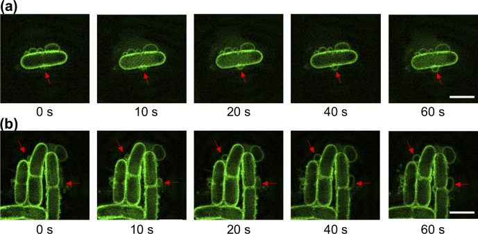
Fig. 4.
Phage infection results in MV blebbing in the absence of cell lysis. Time-lapse image sequence of membrane blebbing in E. coli MG1655 treated with (a) T4 phage or (b) T7 phage. Red arrows indicate site of MV blebs on cell. The membrane dye FM1-43X (green) was used for membrane staining. The phage buffer control in this experiment was visually identical to Fig. 3b. Images were acquired using 3D-SIM from two (t4) or four (t7) independent experiments and are representative of 28 (t4) or 99 (t7) cells with MV blebs. Scale bar is 2 µm. The full time-lapse image series for (a, b) are presented in Movies S7–S8.

