Fig. 5.
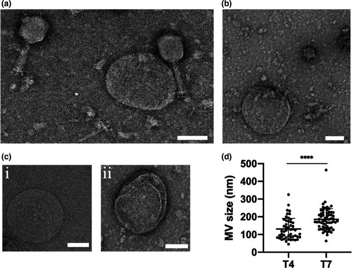
Phage infection of E. coli produces a range of different kinds of MVs within phage lysates. Representative TEM images of (a) T4 lysate with MV and cell debris, (b) T7 lysate with MVs and cell debris. (c) Representative images of different forms of MVs observed within both T4 and T7 lysates: (i) single membrane layer or (ii) multiple membrane layer MVs. (d) MVs sizes within T4 (50 MVs analysed) and T7 (62 MVs analysed) lysates. Data are presented as the mean±sd (****P <0.0001, unpaired t-test with Welch’s correction). For (a, b) images are representative of 49 (T4 lysate) or 61 (T7 lysate) random fields of view imaged in three independent experiments. Scale bar is 100 nm.

