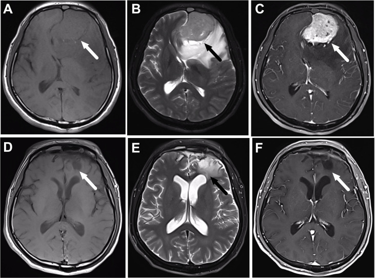
Figure 1.
A 56-year-old male patient with olfactory neuroblastoma of Kadish Stage C underwent surgical resection followed by chemoradiotherapy. (A–C) are the magnetic resonance (MR) images before treatment, while (D–F) are the corresponding MR images after the completion of treatment. (A) Axial T1-weighted MR imaging reveals an isointense mass in the frontal lobe (arrow). The mass has moderate signal intensity on T2-weighted MR images (B), and shows heterogeneous enhancement on the post-contrast T1-weighted MR images (C). (D–F) show that the tumor totally disappears after the completion of treatment, and there is still edema of the frontal lobe (arrow).

