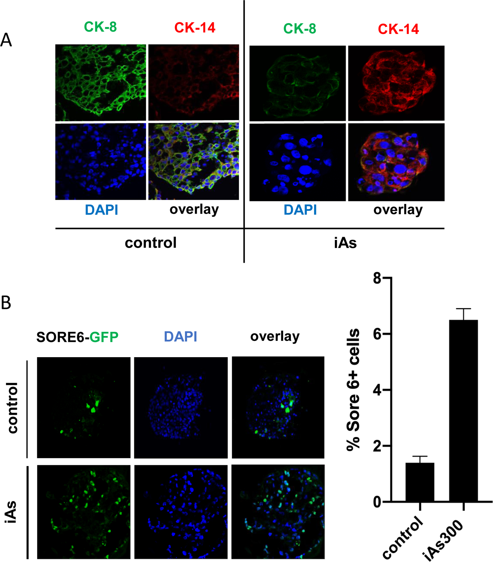
Figure 4. Analysis of luminal (CK8) and basal (CK5) markers as well as cancer stem cells in organoids of MCF-7 cells grown in 3D.

(A) MCF-7 cells (either parental or cells exposed to iAs for 180 days) were grown as organoids in 5% matrigel™. After 21 days, organoids were harvested, fixated with paraformaldehyde and stained for the luminal epithelial cell marker (CK8, shown in green) or basal cell marker (CK5, shown in red). (B) MCF-7 or MCF-7-iAs-180 were transfected with a genetically encoded construct encompassing a Sox2/Oct4 responsive element (SORE6) upstream of GFP. This “stemness” reporter called SORE6-GFP indicated stem cells. Cells were, then platted under low adherence conditions and grown into organoids as in A. Cells displaying GFP positivity were counted using an inverted fluorescence microscope. (C) Quantification of results shown in (B) displayed as averages ± SEM of at least three independent biological replicates. **p < 0.01.
