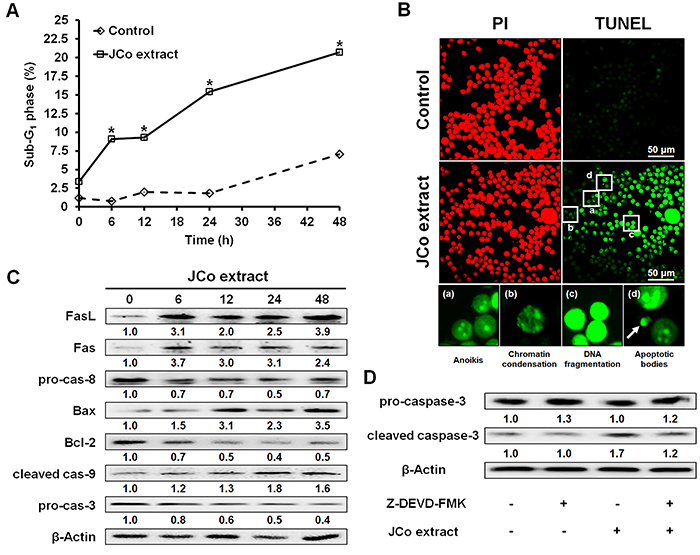
Figure 4. Effects of Juniperus communis (JCo) extract on the extrinsic and intrinsic apoptotic pathways in HT-29 cells. A, The percentage of SubG1 phase cells after JCo extract treatment was analyzed by flow cytometry. Data are reported as means±SD. *P<0.05 vs control (ANOVA). B, Cell apoptosis was determined after treatment with 65 μg/mL JCo extract for 48 h by TUNEL assay (scale bar 50 μm). The apoptotic morphologies included anoikis, chromatin condensation, DNA fragmentation, and the appearance of apoptotic bodies (arrow). C, The protein expression levels of the components of the extrinsic and intrinsic apoptotic pathways in JCo extract-treated cells were analyzed by western blotting. D, HT-29 cells pretreated with 1 μM Z-DEVD-FMK (caspase-3 inhibitor) for 2 h were treated with 65 μg/mL JCo extract for 24 h, and caspase-3 activation was determined by western blotting.

