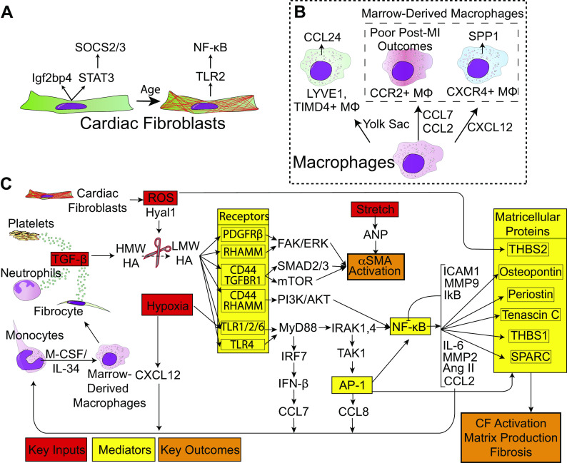
Figure 3.
Model for cellular and molecular changes with age and infarction. A: age-related transcriptional differences in fibroblasts. As fibroblasts progress through development, they upregulate TLR2 expression and downregulate STAT3 and Igf2bp3. B: macrophage composition of the heart shifts from CCL24-producing YS lineage cells to two ontogenies of BMDMs: CXCR4+ and CCR2+. C: proposed molecular mechanism for nonregenerative cardiac fibroblasts. MI in postnatal day 8 hearts generate inflammatory ligands (red) to a greater extent than postnatal day 1 hearts, which are sensed to a greater extent by TLR2, and ultimately result in NF-κB and AP-1 activation (yellow). Lastly, the signal propagation results in excess matricellular protein and ECM deposition (orange). Signaling from receptors and continuing to the right is believed to occur in fibroblasts. AP-1, activator protein-1; BMDM, bone marrow-derived macrophages; ECM, extracellular matrix; MI, myocardial infarction; NF, necrosis factor; TLR, Toll-like receptors.

