. 2021 Jul 8;10:e69544. doi: 10.7554/eLife.69544

Figure 2. DNA repair synthesis in alkylated and undamaged control plasmid DNA in NPE.

(A) Outline of the spot assay. Plasmids were incubated in nuclear extracts supplemented with α32P-dATP; at various time points, an aliquot of the reaction mixture was spotted on DEAE paper (see 'Materials and methods'). The dot blot is shown for the sake of illustration only. (B) Plasmid DNA pBR322 (4.3 kb) samples, modified to a similar extent with -MMS, -MNU and -ENU, were incubated in nucleoplasmic extracts (NPE) supplemented with α32P-dATP at room temperature; incorporation of radioactivity was monitored as a function of time using the spot assay described above (A). Undamaged plasmid DNA0 was used as a control. At each time point, the average values and standard deviation from three independent experiments were plotted. The y-axis represents DNA repair synthesis expressed as a fraction of input plasmid replication (i.e., 10% means that the observed extent of repair synthesis is equivalent to 10% of input plasmid replication). This value was determined knowing the average concentration of dATP in the extract (50 μM) and the amount of added α32P-dATP. (C) N-methyl-N-nitrosourea (MMS)- and N-methyl-N-nitrosourea (MNU)-treated plasmids were incubated in NPE, supplemented or not, by aphidicolin (150 μM final). After 1 hr of incubation, plasmids were purified and analyzed by agarose gel electrophoresis under neutral loading conditions. The gel was imaged by fluorescence (left: ethidium bromide image) and by autoradiography (right: 32P image). The number below each lane indicates the total amount of signals per lane (expressed in arbitrary units [AU]). Aphidicolin treatment decreases incorporation into MNU-treated plasmid close to fourfold, while it affected incorporation into MMS-treated plasmid only 1.6-fold. (D) Samples as in (C). Gel loading is performed under alkaline conditions to denature DNA before entering the neutral agarose gel, allowing single-stranded nicks present in DNA to be revealed. The number below each lane indicates the amount of signals per lane (AU).

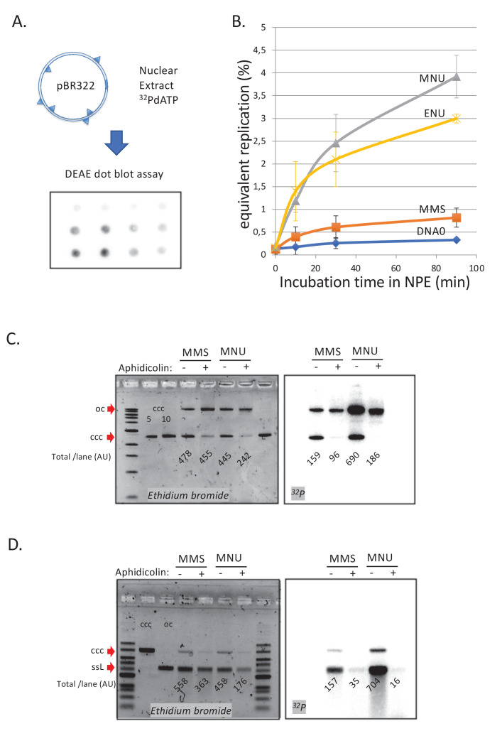
Figure 2.

Figure 2—figure supplement 1. Involvement of mismatch repair in repair synthesis and effect of aphidicolin.

Figure 2—figure supplement 1.

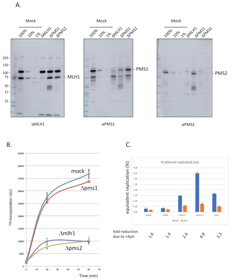
(A) Analysis by western blotting (WB) of the nucleoplasmic extracts (NPE) depleted for mismatch repair (MMR) proteins. Antibodies against Mlh1, Pms2, and Pms1 were used as previously described (Kato et al., 2017; Kawasoe et al., 2016). Extracts were depleted, with the respective antibodies, for three rounds at 4°C at a ratio bead: antibody: extract = 1:3:5. (B) Depletion experiments show that α32P-dATP incorporation into N-methyl-N-nitrosourea (MNU) plasmid is mediated by mismatch repair. NPE was depleted by antibodies against Pms1, Pms2, Mlh1, or mock depleted. Upon incubation at room temperature in different NPE, incorporation of α32P-dATP in MNU plasmid was monitored by the spot assay as a function of time. At each time point, the average values and standard deviations from two independent experiments were plotted. As expected from previous data (Figure 2B), robust incorporation was observed for MNU plasmid incubated in mock-depleted extracts. In contrast, radioactive dATP incorporation was severely reduced when the plasmid was incubated in extracts depleted with antibodies against Mlh1 or Pms2. These data strongly suggest that the incorporation seen in mock-depleted NPE is mediated by MMR, as both Mlh1 and Pms2 assemble into the functional MMR complex MutLα. In contrast, depletion with antibodies against Pms1 did not affect incorporation kinetics as Pms1 is reported not to function in MMR (Jiricny, 2006). (C) Effect of aphidicolin: alkylated plasmids (pEL97: 11.3 kb) were incubated in NPE supplemented or not by aphidicolin (150 μM final) in the presence of 32P-dATP for 1 hr at room temperature (RT). Analysis was performed by spot assay as described in 'Materials and methods'. The y-axis represents the percentage of radioactive DNA synthesis expressed in % of full plasmid replication. Addition of aphidicolin to NPE (150 μM final) more severely reduces incorporation in MNU+, MNU++, and ENU plasmids, compared to control and methyl-methane sulfonate (MMS) plasmids. Fold reduction in incorporation under aphidicolin conditions is shown.
Figure 2—figure supplement 2. Repair synthesis in HSS extracts.

Figure 2—figure supplement 2.

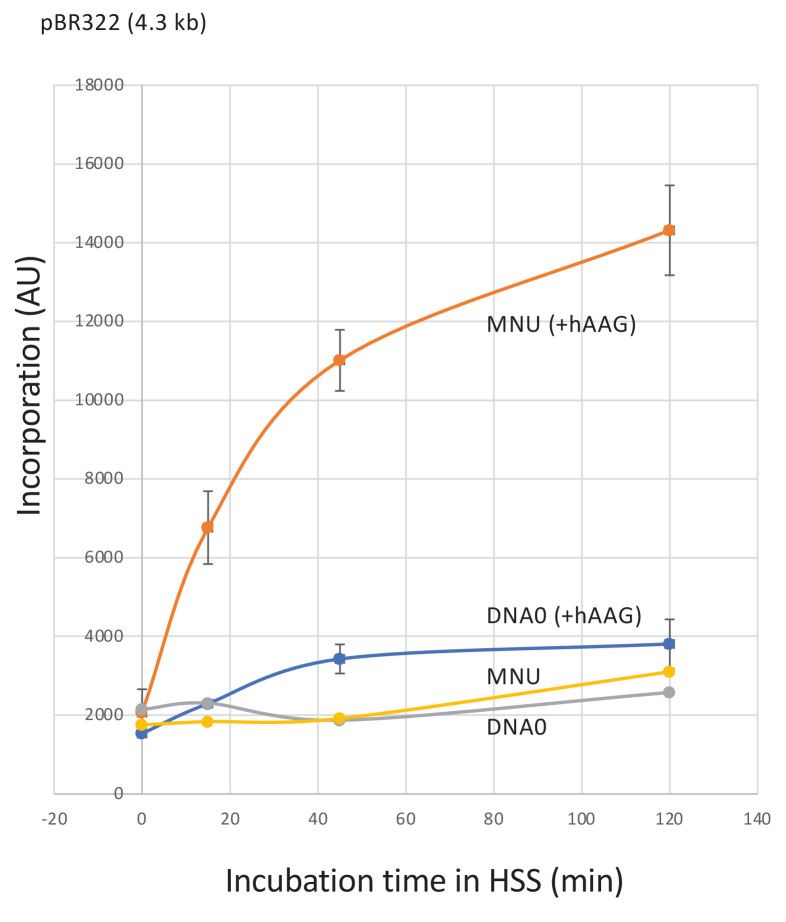
Plasmid treated with N-methyl-N-nitrosourea (MNU) (≈1N-alkyl adduct/500nt) was incubated in high-speed supernatant (HSS) extracts in the presence of α32P-dATP; repair synthesis was monitored at room temperature as a function of time using the spot assay. Both DNA0 and MNU plasmids exhibited background incorporation levels. Robust unscheduled DNA synthesis (UDS) was observed in MNU plasmid upon addition of hAAG glycosylase (150 nM). Average values and standard deviations from two independent experiments were plotted.