. 2021 Jul 8;10:e69544. doi: 10.7554/eLife.69544

Figure 4. Double-strand breaks occur in MNU-treated plasmids during incubation in extracts.

(A) Analysis by agarose gel electrophoresis (AGE) of alkylated plasmids (pEL97: 11.3 kb) incubated in nucleoplasmic extracts (NPE) in the presence of α32P-dATP. Plasmid pEL97 was treated with methyl-methane sulfonate (MMS), N-methyl-N-nitrosourea (MNU)+, and ENU as to introduce ≈ one alkylation event, on average, every 500 nt. For MNU, a plasmid with twice the level of alkylation (MNU++, one lesion every 250 nt) was also produced (Figure 4—figure supplement 1). Alkylation of these plasmids essentially not affected their migration on agarose gels (Figure 4—figure supplement 2A). After 2 hr of incubation, the reaction was stopped and a known amount of pBR322 (10 ng) plasmid was added as an internal standard. Ethidium bromide image: in different lanes, the internal standard band, pBR (covalently closed circular [ccc]), appears to be of similar intensity (1158 +/- 95 arbitrary units [AU]), assessing reproducible DNA extraction. For the alkylated plasmids, incubation in NPE led to massive conversion from ccc to relaxed plasmids. 32P image: little incorporation of 32P-dATP is seen in DNA0 and in MMS-treated plasmids compared to MNU- and ENU-treated plasmids as shown by the relative incorporation levels normalized to one for untreated plasmid (DNA0). As expected, the MNU++ sample exhibits about twice the amount of incorporated radioactivity compared to MNU+. In both ethidium bromide and 32P images, a small amount of linear plasmid is seen mostly in the MNU++ sample. This band is also visible in the MNU+ and ENU lanes although at a weaker intensity. (B) Quadratic dose-response for double-strand break (DSB) formation. When the % of linear form (linear/(linear + oc)) is plotted as a function of the square dose of MNU (mM2) for untreated, MNU+, and MNU++ plasmids, we observed a straight line (y = 1.4173x - 0.0288; R² = 0.9999).

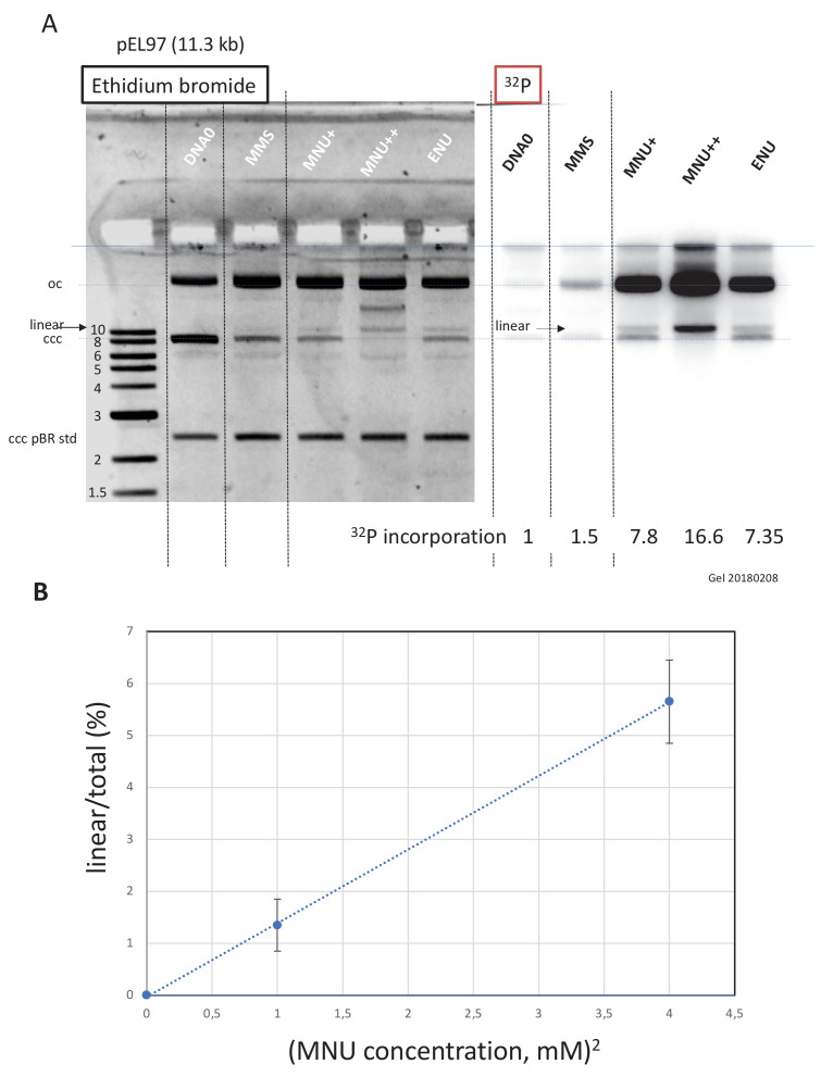
Figure 4.

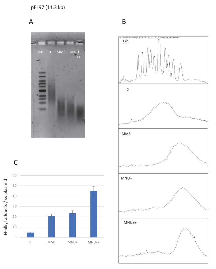
Figure 4—figure supplement 1. Estimation of N-alkylation levels of modification by MMS and MNU.

Figure 4—figure supplement 1.

(A) Alkaline fragmentation of alkylated plasmid (pEL97, 11.3 kb) DNA is analyzed by agarose gel electrophoresis. (B) Densitometry of the ethidium bromide-stained agarose gel. (C) Estimation of the average number of alkali cleavage sites per plasmid strand. The data reveal that the average number of cleavage sites per plasmid strand is 21–23 for both methyl-methane sulfonate (MMS) and N-methyl-N-nitrosourea (MNU)+ reaction conditions (the average distance between two sites is ≈510 nt). For MNU++, the average distance between two cleavage sites is ≈254 nt.
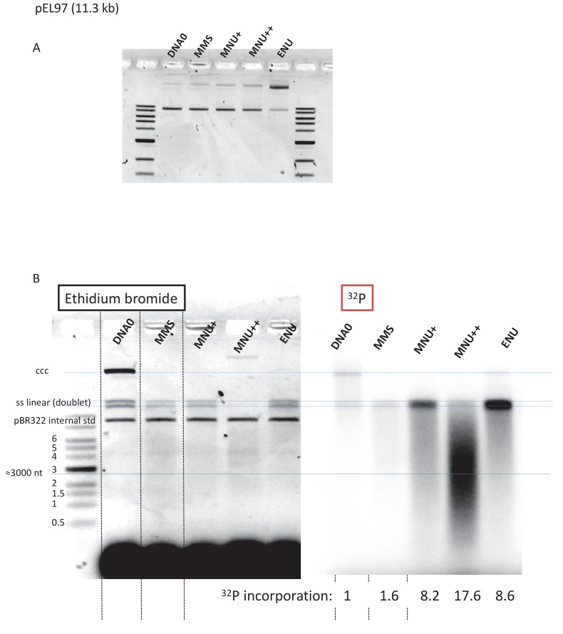
Figure 4—figure supplement 2. Fragmentation of alkylated plasmid as analyzed on AGE loaded under alkaline conditions.

Figure 4—figure supplement 2.

(A) Alkylation did not affect plasmid topology except for ENU treatment that increases relaxation as seen in the agarose gel electrophoresis (AGE) image. (B) Analysis by AGE, under alkaline loading conditions, of alkylated plasmids (pEL97: 11.3 kb) incubated in nucleoplasmic extracts (NPE) in the presence of α32P-dATP (same samples as in Figure 4A). Loading under alkaline conditions allowed single-stranded nicks to be revealed. In the different lanes, an internal standard band, pBR (covalently closed circular [ccc]), was introduced before the extraction procedure in order to assess the consistent recovery of DNA during extraction (as seen by ethidium bromide staining). For the alkylated plasmids, incubation in NPE led to massive conversion of plasmid DNA into a smear with a low amount of DNA present as a single-stranded linear band (doublet). For the MNU++ sample, no linear single-stranded DNA (ssDNA) was seen; all DNA was converted into a smear. The smearing can best be seen in the 32P image. We suggest that the ssDNA fragments that form the smear arise between a gap caused by a mismatch repair (MMR) event at an O6mG site and an uncompleted base excision repair (BER) event at a neighboring lesion. It cannot be excluded that some fragments result from MMR attempts at two O6mG lesions. The intense 32P labeling of the fragments in the smear results from incorporation of up to several hundred nt during a single MMR repair synthesis. Relative 32P incorporation normalized to one for DNA0 is indicated below the 32P image.