Fig. 5.

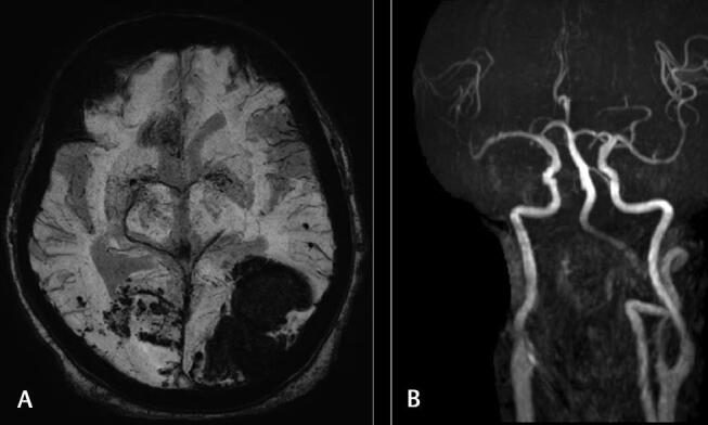
( A ) Susceptibility-weighted imaging (SWI) images showing hemorrhagic areas in both parieto-occipital lobes. ( B ) Magnetic resonance angiography (MRA) showing hypoplastic right vertebral artery on MRA.

( A ) Susceptibility-weighted imaging (SWI) images showing hemorrhagic areas in both parieto-occipital lobes. ( B ) Magnetic resonance angiography (MRA) showing hypoplastic right vertebral artery on MRA.