
Fig. 2.
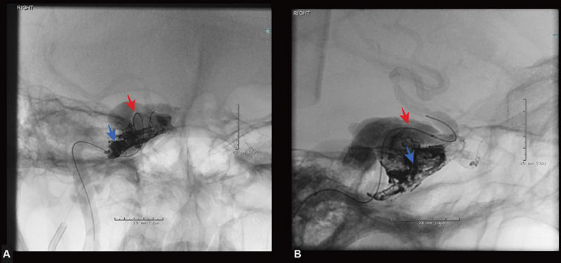
Periprocedural images of Onyx embolization of right carotid-cavernous fistula. ( A ) Anteroposterior view of right cavernous sinus angiogram showing the balloon in the right cavernous internal carotid artery (red arrow) and an Apollo catheter in the right cavernous sinus through the inferior petrosal vein (blue arrow). ( B ) Lateral view of the right cavernous sinus showing a deflated balloon catheter in the right carotid-cavernous artery and Onyx cast in the right cavernous sinus.