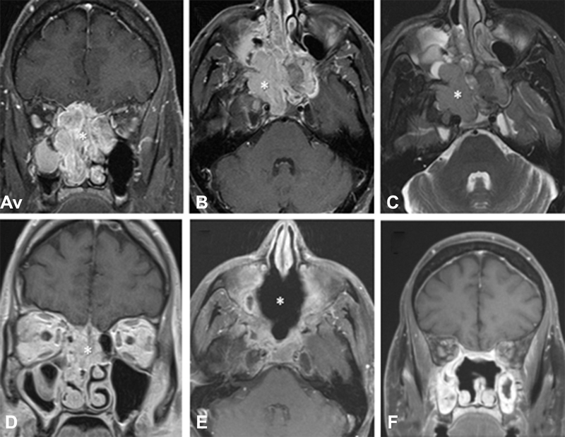
Fig. 3.

Magnetic resonance imaging of a 44-year-old male presenting with esthesioneuroblastoma initially deemed unresectable prior to induction therapy (patient 1). ( A ) Coronal T1-weighted postgadolinium image demonstrating a large sinonasal mass (*). ( B ) Corresponding axial T1-weighted gadolinium-enhanced image. ( C ) Corresponding axial T2-weighted image. ( D ) Coronal T1-weighted postgadolinium image status postinduction chemotherapy with two cycles of cisplatin, doxorubicin, and vincristine, followed by two cycles of etoposide and cisplatin and then two cycles of etoposide and cisplatin with concurrent 6,480 cGy of external beam radiotherapy in 36 fractions. This image was taken immediately prior to surgery, which was achieved in a gross total fashion with negative microscopic margins. ( E ) 4-month postoperative axial T1-weighted gadolinium enhanced imaging demonstrated no evidence for residual or recurrent disease. ( F ) Corresponding coronal T1-weighted gadolinium-enhanced image.
